Biology 111
More Cells!
Calibrating the microscope:
You cannot tell, in absolute terms, how big any specimen under your
microscope is without calibrating the microscope first. You will always have an
ocular micrometer in one of your oculars, but using this alone gives only a
relative indication of size. You must use a stage micrometer to calibrate
the scope, and in doing so, you will find out:
What does one ocular unit
represent in absolute units?
You can then use the ratio you found
to calculate the size of specimens. You must calibrate the scope at each
magnification that you intend to use.
Something about calibration, or its
downstream activities (calculating size, creating a scale bar) is always
on the lab exam.
Plant tissues: We looked only
at a vascular bundle from the stalk of Swiss Chard (Beta vulgaris). The
vascular bundle is made up of a cap of smaller tubes known as phloem.
These tubes carry the plant's food (what is the plant's food, where does it come
from?). The larger, thicker-walled tubes are the xylem vessels; these transport
water. The parenchyma cells are the larger, blockier cells which surround and
support the vascular bundles.
| Cross Section |
 |
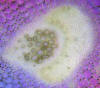 |
Try as I might, I couldn't get a
decent picture of a vascular bundle, so I've provided links to two
shots. In the first picture, the massive
squarish patch of large white cells is made up of parenchyma. The xylem
borders this (stained red), the phloem is the beige (unstained),
crescent-shaped patch adjacent to the xylem. This photo is not from
celery or Swiss chard, thought the bundles are representative.
In the second picture (from celery), the parenchyma is
the mass of purple-staining cells surrounding the bundle. Xylem tubes
are large and thick-walled (the cluster on the left), the phloem are
smaller and form into an arc-shaped bundle. |
| Longitudinal Section |
 |
 |
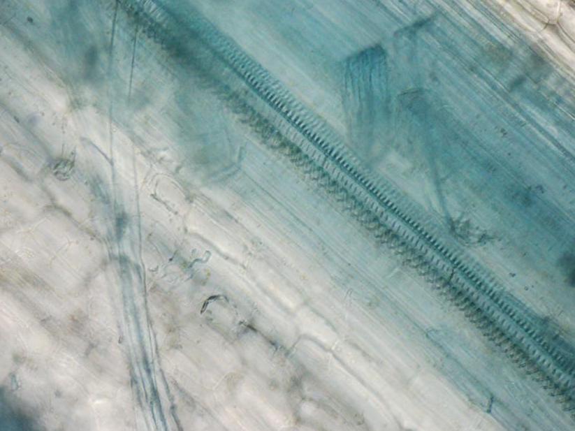
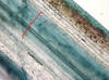
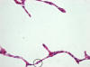
So I was a bit better at the longitudinal
sections. In the first slide, the red bracket denotes one vascular
bundle. The second slide is at higher mag, and shows the curly nature of
the xylem wall. |
Animal tissues: The study of
tissues is known as histology (this term applies to
animals and plants). There are four basic tissue types in animals, we
looked only at epithelial tissues. Epithelium lines cavities or covers an organ;
ergo, look for these tissues next to some type of space. Recall that the word
simple, when used in a histological sense, means one layer; in
contrast, stratified refers to an epithelium of more than one layer of cells.
| Specimen |
broad view |
zoomed in |
notes |
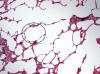
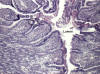
| simple squamous epithelium - lung |
 |
 |
Squamous - sounds like squished.
Good reason for that. In the first slide, the ellipse shows one
alveolus. Note in the second slide how a squamous cell bulges in the
position held by the nucleus. |
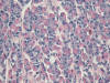
| simple cuboidal epithelium - kidney |
|
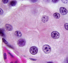 |
This is from one of the urine-producing tubes known as
a nephron. |
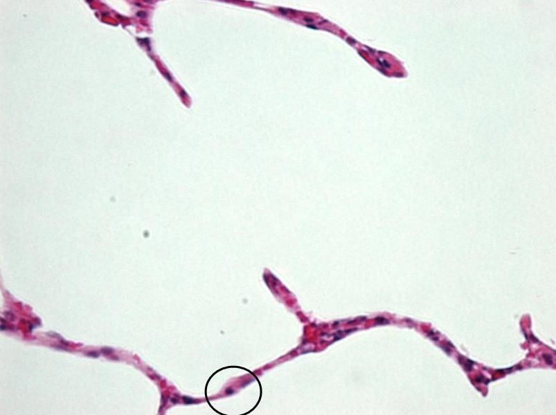
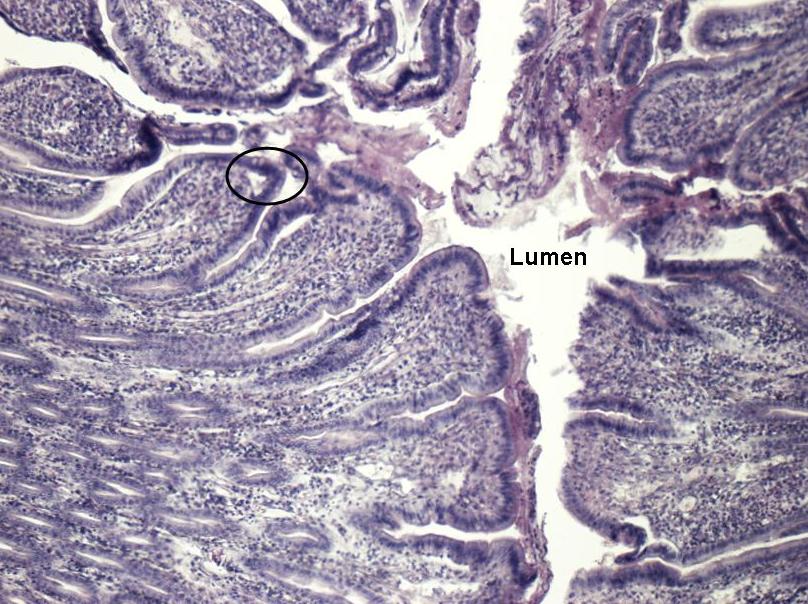
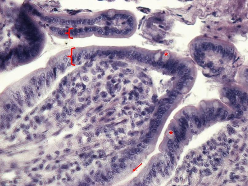
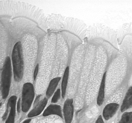
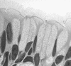
| simple columnar epithelium - small
intestine |
 |
 |
Look for goblet (*) and absorptive cells. In the
latter, note that the nuclei are more or less all at one level. The open
brackets denote the single layer (simple) of cells making up this
tissue. The arrow points to the brush border. |
| pseudostratified ciliated columnar
epithelium - trachea |
|
 |
What is it about this simple tissue that gives it
a stratified appearance? |
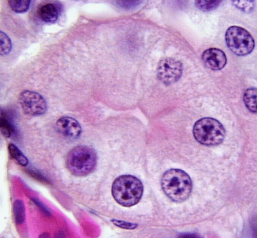
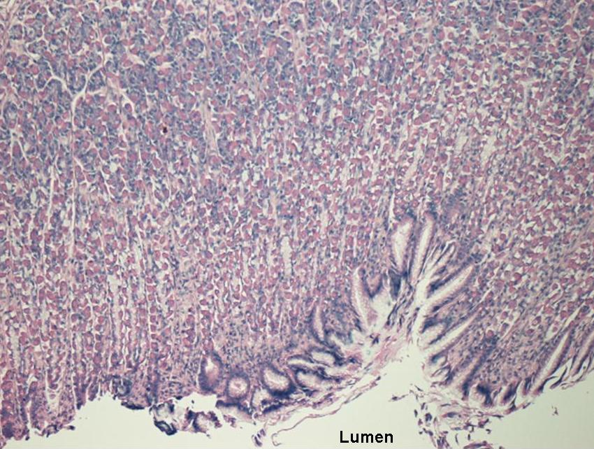
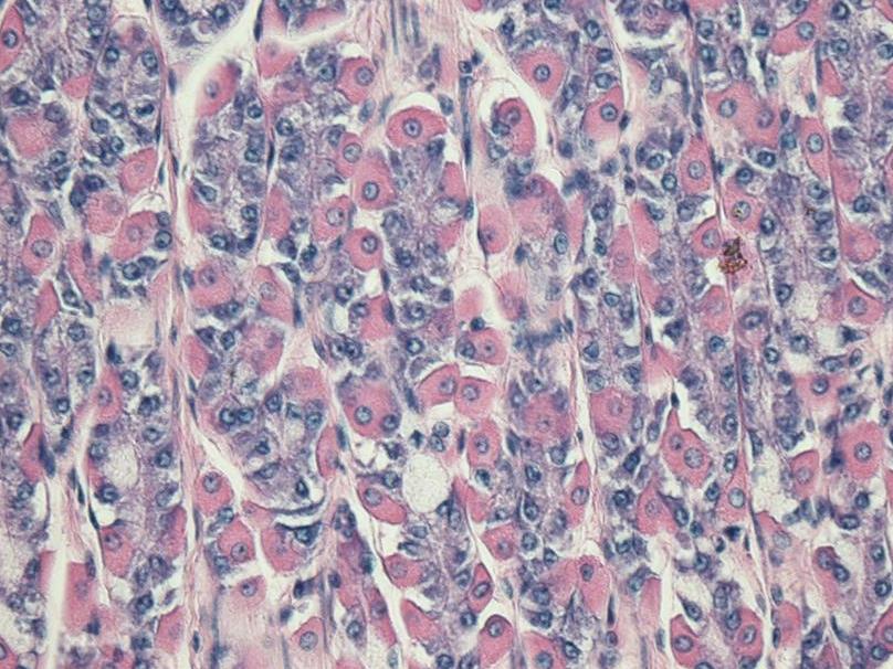
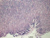
| fundic area of the stomach |
 |
 |
Look for both parietal and chief cells. |

 |
R.F. Lauff
Department of Biology
St. Francis Xavier University
Antigonish, NS Canada B2G 2W5
|