Laboratory Photographs
|
On this page I will put some photos from lab specimens, with
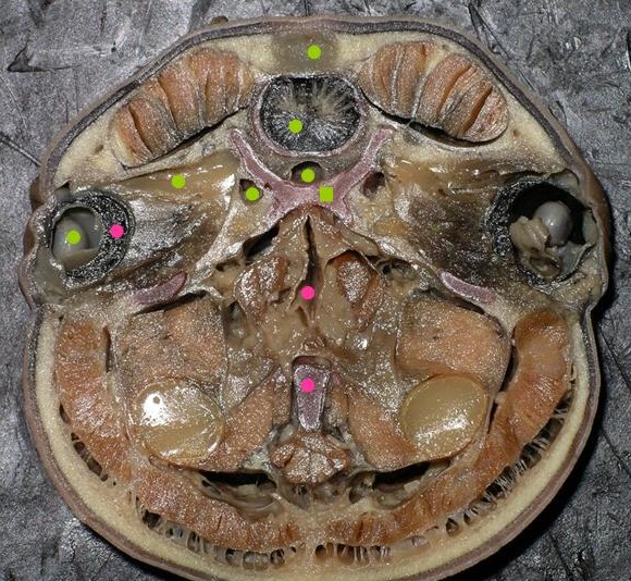
clarifications if necessary. For instance, the photo at the right is of what is illustrated in Figure 3.8 (a) of your lab manuals. You should be able to identify all structures labelled with dots or squares (click on the image for a larger view). The green dots are labelled in figure 3.8 (a); the green square above the upper pink dot is correctly placed on the cranial cartilage. The pink dots are structures not labelled in 3.8 (a), but they are labelled on figure 3.6 (c) - what are they? |
|