The Biology Department and the Human Nutrition Department share the J. Bruce Brown Hall, a four storey structure located on the western side of Saint Francis Xavier University Campus.
J. Bruce Brown Hall's location on campus can be seen in the aerial photo of Campus, or can be found on the electronic map.
The Biology department is housed in a modern, air-conditioned building shared with the department of Nutrition and Consumer Studies and equipped with all modern services including a standby generator. It is well provided with both teaching and laboratory space.
Photosynthesis Lab | Radioisotope Lab | Electron Microscope Laboratory
Biomechanics Research Laboratory | Bacterial Pathogenics Laboratory
Herbarium | Light Microscope Facility | Animal Holding Facility
Greenhouse | Controlled Environment Rooms and Chambers
Fully Equipped Service Room | Darkroom
Photosynthesis Lab - Research in this laboratory is focused upon the role of active C02 and HC03- transport in cyanobacteria, which are of crucial importance to global photosynthetic C02 fixation. Work by Tony Miller and others has shown convincingly that C02 fixation in cyanobacteria requires active transport of C02 and HC03- into the cells. Inorganic carbon accumulated in cells as a result of this causes increased rates of linear electron flow and chlorophyll a fluorescence quenching.
|
The laboratory contains:
|
|
Radioisotope Laboratory - This facility, renovated to AECB specifications in 1989, comprises two rooms, one 7.5 x 8.5 m and a smaller 4 x 5 m. The former is for physiological experiments that use non-volatile radioisotopes and includes scintillation counter (Packard 2000CA purchased in 1988) membrane chambers, current-voltage clamps, gas-mixing pump, chloridometer and atomic emission/absorption spectrophotometer. The small room has a fume hood for working with volatile isotopes. Newly acquired (1991-93) patch clamp facilities (Axopatch 1C and suite of related instruments) and cell culture containment hood (Forma) are used to study ion channels. There are also complete facilities for intracellular recordings of voltage, ions (liquid ion exchange microelectrodes), and pH (using BCECF microspectro-fluorometery) used in studies of membrane transport biology. Respiration and acid-base balance studies are aided by an automated C02 analyzer (Corning), gas mixing pump (Wosthoff), autotitration suite (Radiometer) and pH/pIon meters (Orion). Intracellular recordings are aided by vibration-damped tables (Technical Mfg. Corp.), microelectrode amplifier (WPI KS-700), high impedance microelectrode amplifier (WPI FD223), storage oscilloscope, stimulator and micropositioner (Haer) with a 4 channel oscillographic recorder (Hewlett Packard). Computing capabilities are provided currently by UNIX operating system running on a Tandy 4000 with a peripheral terminal for multi-user access to custom-designed C-language data reduction, bibliographic statistical and graphics routines. A system upgrade is imminent. Membrane transport library comprises 3200 reprints accessible through the UNIX terminals.
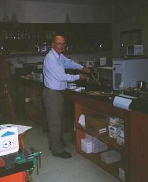
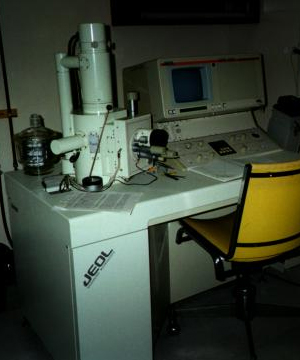
Electron Microscope Laboratory - Acomplete electron microscope facility housed on the second floor (Rm. 225) is fully operational with the recent acquisition of a new JEOL 5300 Scanning Electron Microscope (NSERC grant to Buckland-Nicks, Garbary and Melchin) with Image Analysis system and high resolution video printer. The scanning EM is housed in a room adjacent to a Philips 300 Transmission Electron Microscope which was purchased by Buckland-Nicks from McGill in 1991. Separating the two electron microscopes is a small darkroom for developing negatives. The following auxiliary equipment is available to users of the facility:
- Two Ultramicrotomes;
- Sputter Coater;
- Critical Point Dryer;
- LKB glass knifemaker.
 |
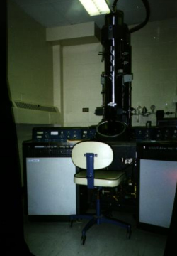 |
| TEM | SEM |
Since setting up our facility in 1991, electron microscopy has been an important element in research projects of six of our nine faculty members and three faculty from other science departments.
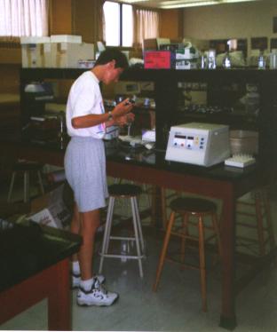
Biomechanics Research Laboratory - The laboratory contains two separate data acquisition facilities, each controlled by LabView. All mechanical testing are done on these units. One system consists of integrated instruments to measure the dynamic physical properties of arteries, and the second unit is centered around a Cambridge Technology Muscle Lever System (2 units are in the facility).
Each unit has associated equipment, including split-screen video microscopy, and a Video Dimension Analyzer, computer controlled water baths, pumps, etc. Four personal computers are located in the room, two for the data acquisition facilities, and two for data analysis, processing, etc. Three of the computers are connected to the St.F.X. local area network. There is also a IBM Xterminal in the lab. An extensive biomechanics library (almost 1400 references) is contained in a database that is accessible on one of the computers. The laboratory also contains a Leitz Polarizing photomicroscope with capability of making quantitative measurements of birefringence.
Bacterial Pathogenics Laboratory - This laboratory meets Level II containment requirements and uses molecular biology techniques and modern methods in cryo-electron microscopy to examine interactions between bacterial pathogens and eukaryotic host cells.
|
Facilities include:
|
|
-
A liquid nitrogen cryo-storage unit for long term storage of eukaryotic cell lines and equipment for the ultra-rapid freezing and processing of specimens for freeze-substitution electron microscopy are also present.
-
An ultramicrotome has recently been added to this lab.
-
Associated with this lab is a tissue culture facility containing a laminar flow hood and two CO-2 incubators for culture and eukaryotic cells.
Herbarium - The herbarium consists of about 3000 herbarium specimens divided almost equally into collections of marine algae and vascular plants. The herbarium is utilized as a resource in teaching both undergraduate and graduate courses, and is a repository for collections related to several ongoing research programs in the department. The collections are stored in four metal and five wooden cabinets, which are housed in a teaching laboratory restricted to advance d courses in plant taxonomy and phycology. Additional herbarium facilities include a small collection of liquid preserved specimens, several stereomicroscopes with stands suitable for examining herbarium specimens, a research quality photo- microscope an d a small library of appropriate identification manuals.
Light Microscope Facility - A Zeiss Photomicroscope III equipped for bright field, fluorescence and Nomarski DIC microscopy is located in room 410. This departmental facility enables accurate microspectrophotometry.
Also available for observation of cells in cultures is an inverted Zeiss photomicroscope. Room 411 houses a Wild M420 macroscope equipped with a macrozoom lens and a fibre-optic bright field/darkfield transmitted light base, purchased in 1996 for high resolution microscopy and microphotography of large biological specimens at intermediate magnifications (up to 87 X).
Animal Holding Facility - small unit (2250 ft2) with an office, cleaning and storage area, five small animal holding rooms, and a large aquatic room. Research is carried out in the facility's two walk in controlled-environment chambers.
Greenhouse - located on the fourth floor of the Biology building the greenhouse (10 x 7 m) provides horticultural facilities required for both teaching and research. This is organized and run by the Manager of Animal and Plant Care - Anne Louise MacDonald.
Controlled Environment Rooms and Chambers - Six controlled environment chambers (temp. and light) and six constant environment rooms are located in various labs around the department. These facilities are maintained by the university.
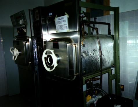
Fully Equipped Service Room - On the second floor the department maintains a service room including a still, crushed ice machine, laboratory glassware dishwasher, pipet washer, drying oven and two autoclaves.
Darkroom - A fully equipped professional darkroom is located on the fourth floor of the department for black and white processing in a variety of formats. Beseler and Durst enlargers are available for general use.