|
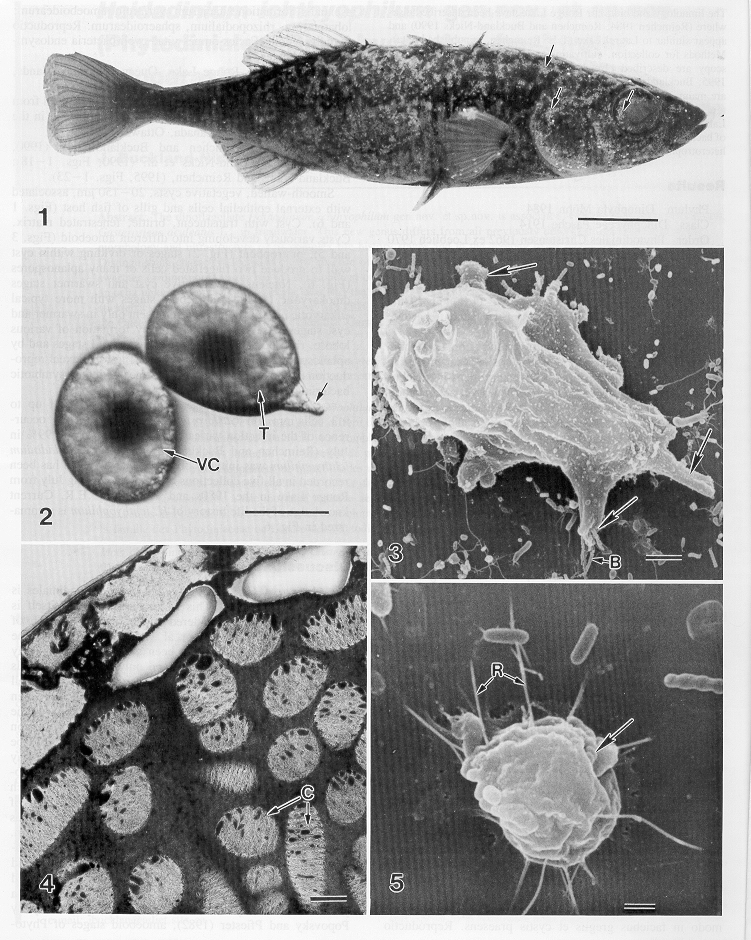
1
|
Spine-reduced
three spine stickleback, Gasterosteus aculeatus showing heavy infection
with vegetative cysts of Haidadinium icthyophilum. Arrows indicate
concentrations of cysts on dorsal surface, operculum and eye. Scale bar
= 1cm. |
|
2
|
Vegetative
cyst (VC) of H. icthyophilum forms trophont (T) by extension of stalk-like
organelle (arrow), which attaches to algal filament. Scale bar = 10m m. |
|
3
|
Lobose
amoeba of H. icthyophilum, with several extende lobopodia (arrows) phagocytoses
bacteria (B). Scale bar = 10m m. |
|
4
|
Transmission
electron micrograph of dinokaryotic nucleus of newly formed vegetative
cyst, showing condensed chromosomes (C). Scale bar= 0.5 m m |
|
5
|
Rhizopodial
amoeba with fine rhizopods (R). Note engulfed spiral bacterium (arrow).
Scale bar = 1m m |

