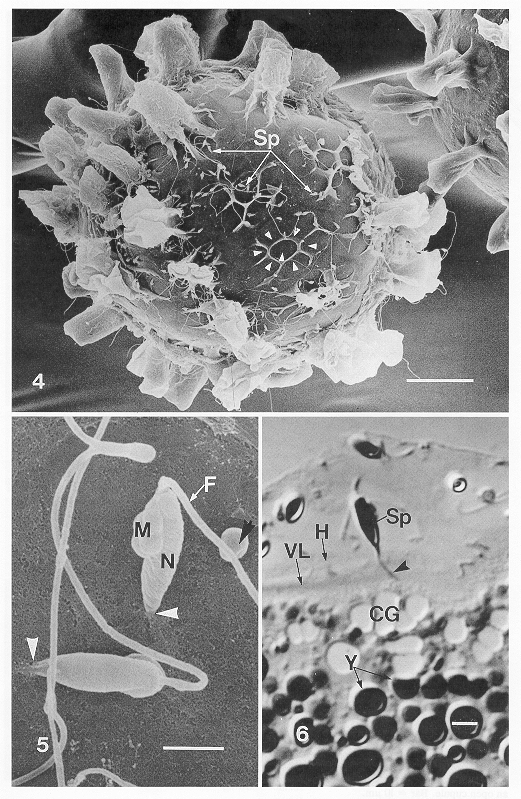
|
4
|
SEM
of zygote with some hull cupules torn away showing channels that partition
them (arrowheads). Note sperm (Sp) penetration at these and other locations
where the hull is exposed. Closed cupules on an adjacent egg did not attract
sperm. Bar = 40m m. |
|
5
|
Part
of figure 4 at higher magnification to show two fertilizing sperm that
have penetrated the hull (white arrowheads) and have not entered through
a micropyle. Note features of sperm such as basal mitochondria (M), main
body of nucleus (N), and flagellum (F). A rounded structure attached to
the flagellum may once have been the lateral mitochondrion (black arrow).
Bar = 2m m. |
|
6
|
Light
micrograph of a 0.5m m section through a fertilizing sperm (Sp), with anterior
filament (arrowhead) penetrating the hull (H) and vitteline layer (VL)
at the base of a cupule. CG, Cortical granules; Y, yolk granules. Bar =
2m m |