|
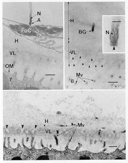
7
|
TEM
of the acrosome (A) of a sperm that did not react after contacting a follicle
cell (FoC) between the hull cupules. Note also nucleus (N) within sperm
anterior filament, hull (H), vitelline layer (VL), and oocyte membrane
(OM). Bar = 0.5m m. |
|
8
|
TEM
through tip of sperm anterior filament in the hull (H). The acrosome has
fired, the apical granule has been exhausted, and the basal granule (BG)
has been exposed. Clear areas (arrowheads) indicate pores through the vitelline
layer (VL). Tips of egg microvilli (Mv) can be seen below the pores in
this rare oblique section through the egg envelopes. Bar = 0.5m m. Inset:
Tip of sperm anterior filament enlarged to show basal granule (arrowhead)
and nucleus (N). Bar = 0.1m m |
|
9
|
TEM
of section perpendicular to the oocyte membrane of an unfertilized egg
showing pores (arrowheads) through the vitelline layer (VL). Also visible
are some microvilli (Mv) that have penetrated the vitelline layer and hull
(H). Bar = 0.5m m. |