|
10
|
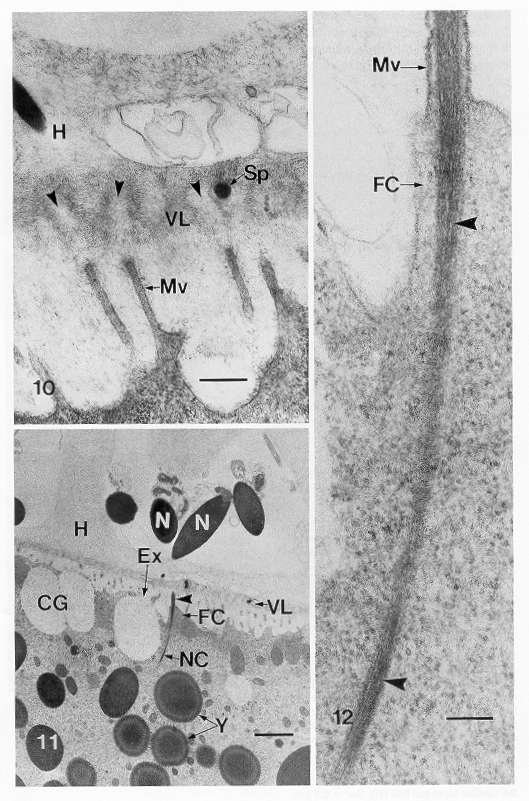
TEM
of a section perpendicular to the oocyte membrane of a zygote illustrating
pores (arrowheads) through the vitelline layer (VL). Each pore leads to
the tip of a single egg microvillus (Mv). Note section of sperm nucleus
(Sp) within one pore. H, hull. Bar = 0.5m m |
|
11
|
TEM
of fertilization. Anterior filament of sperm has fused with a single microvillus
of the oocyte membrane (arrowhead), and nuclear chromatin (NC) is being
injected into the egg. One cortical granule has been released by exocytosis
(Ex), and two others (CG) are adjacent to the egg membrane. Note also the
main body of sperm nucleus (N), hull (H), vitelline layer (VL), apparent
fertilization cone (FC), and yolk granules (Y). Bar = 1 m m. |
|
12
|
Site
of fusion of sperm anterior filament with egg microvillus (Mv), showing
sperm chromatin (arrowheads) entering egg cytoplasm devoid of nuclear membrane.
FC, apparent fertilization cone. Bar = 0.1m m. |

