|
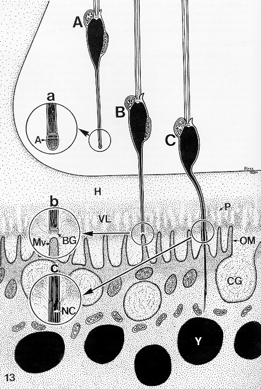
Three
sperm are depicted at the base of a cupule in different stages of penetration.
Sperm A approaches the hull, with acrosome intact (see circle a).
Sperm B has penetrated the hull and located a pore in the vitelline
layer; the acrosome has fired, the apical granule has been exhausted, and
the basal granule is being used up as the tip of the anterior filament
approaches an egg microvillus (see circle b). Sperm C has
fused with an egg microvillus, creating a membranous tube through which
the sperm chromatin is being injected into the egg cytoplasm, devoid of
nuclear membrane (see circle c). Acrosome (A), hull (H), vitelline
layer (VL), pore through vitelline layer (P), basal granule (BG), egg microvillus
(Mv), oocyte membrane (OM), cortical granule (CG), yolk granule (Y), and
nuclear chromatin (NC) are also shown. |