| Specimen | Bright Field | Dark Field | Polarization |
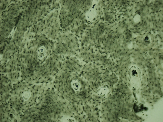
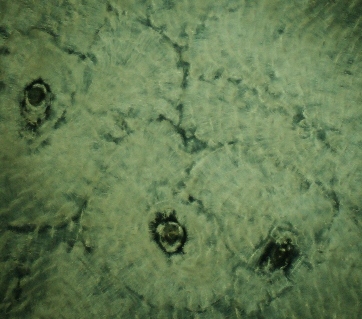
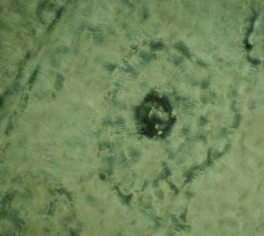
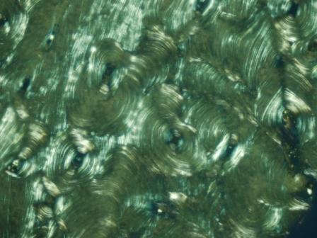
| Bone - Haversian System |
 |
 |
 |
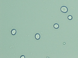
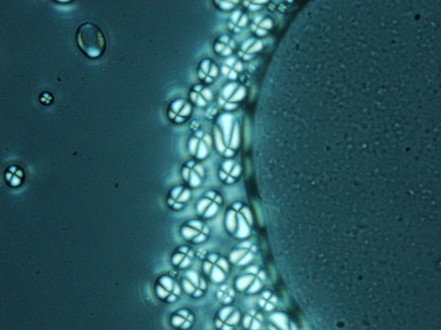
| Starch Granules - unstained |
 |
 |
 |
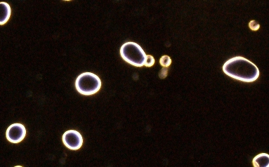
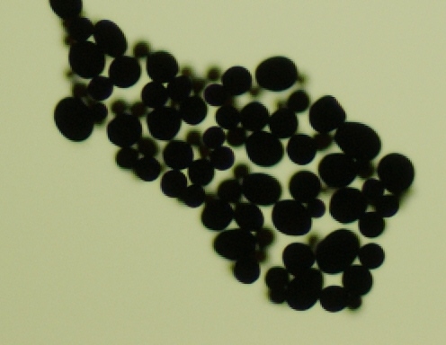
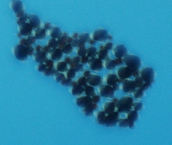
| Starch Granules - stained |
 |
 |
 see
notes see
notes |
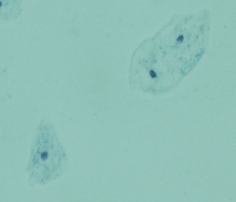
| Cheek Cells |
 |
 |
 |
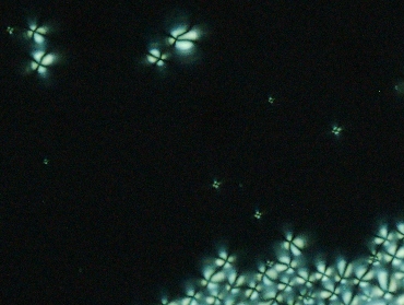
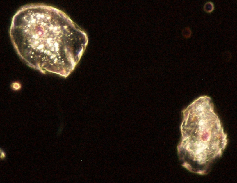
| Live Microorganisms |
Notes: The key to Dark Field is a crystalline structure or halo effect of the specimen; the background is dark, not necessarily blue. The blue background in the image of the stained potato starch granules is the result of looking through polarizing filters...when they are in the cross alignment, the background is back, and so too will be the specimen if it is not birefringent. The stained granules are not birefringent, so I have shown that they are there by not having the filters cross-aligned.