Biology 304
Muscle Lab Equipment Overview
Biology 304
Muscle Lab Equipment Overview
|
|
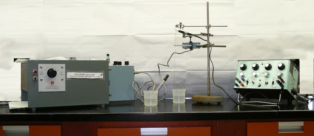
| The big picture. This is how you will see the equipment when you come to lab. |
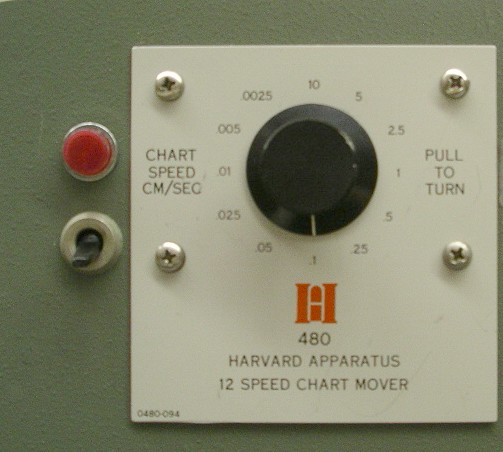 1.
The chart recorder is on the left. It is simply a box with a source of paper and
a motor which moves the paper at a speed that you choose; the paper speed
selector is magnified on the left, the power switch is below the red light. The
muscle contractions are recorded on this paper, and just as importantly, your
observations and procedures (stimulus strength, load, addition of drugs, etc)
are recorded by directly writing on the paper.
1.
The chart recorder is on the left. It is simply a box with a source of paper and
a motor which moves the paper at a speed that you choose; the paper speed
selector is magnified on the left, the power switch is below the red light. The
muscle contractions are recorded on this paper, and just as importantly, your
observations and procedures (stimulus strength, load, addition of drugs, etc)
are recorded by directly writing on the paper.
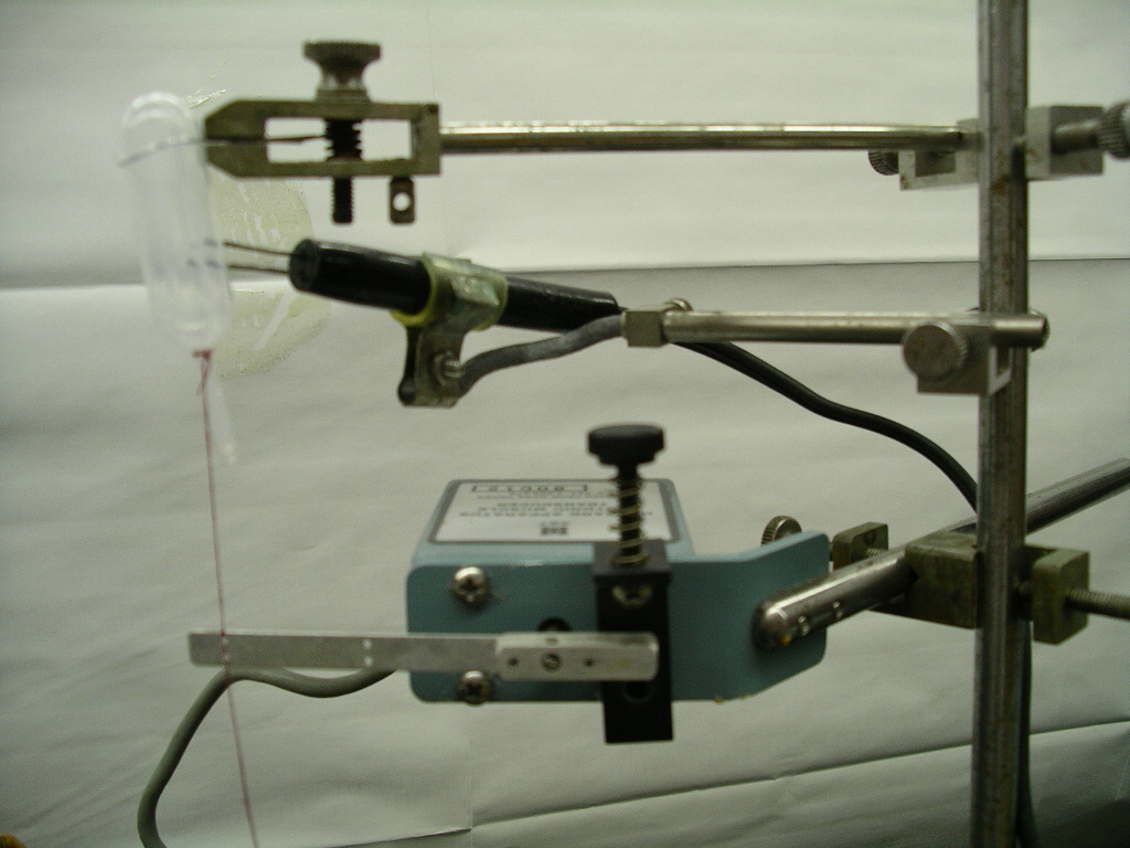 2.
The dissected muscle is hooked up to a transducer whose role is to convert the
muscles movement to an electrical signal proportional to the amount of
movement. The transducer is the lowest piece of equipment on the retort stand in
the photo at the left. The upper piece is the femur clamp, the middle piece is
the electrode.
2.
The dissected muscle is hooked up to a transducer whose role is to convert the
muscles movement to an electrical signal proportional to the amount of
movement. The transducer is the lowest piece of equipment on the retort stand in
the photo at the left. The upper piece is the femur clamp, the middle piece is
the electrode.
![]() 3.
The recorder module is similar to your stereos amplifier both take an
electrical signal, amplify it and provide an output. The recorder module takes
the electrical signal from the transducer, amplifies it and sends that amplified
signal to the pen which then moves proportional to the signal strength. As the
paper from the chart recorder moves, the pen produces a drawing of the muscle
contraction.
3.
The recorder module is similar to your stereos amplifier both take an
electrical signal, amplify it and provide an output. The recorder module takes
the electrical signal from the transducer, amplifies it and sends that amplified
signal to the pen which then moves proportional to the signal strength. As the
paper from the chart recorder moves, the pen produces a drawing of the muscle
contraction.
And if all goes well...you get a tracing produced!
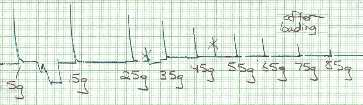
Note that in this example, the students have written down the part of the experiment they are doing "after loading" and have labelled each part of the tracing with the mass tested (e.g. 15 g, 85 g); they have also x'd out mistakes...this way there is no confusion as to what is a muscle contraction and what is an "oops".
 |
Department of Biology St. Francis Xavier University Antigonish, NS Canada B2G 2W5 |